Understanding Cytopathology: Decoding Cellular Health Today
Sep 30, 2026 Diagnostic

Understanding Cytopathology: Decoding Cellular Health Today

An essential guide to cell-based diagnostics, early disease detection, and clinical accuracy.

Cytopathology is a specialized branch of pathology that studies diseases at the cellular level. Unlike histopathology, which examines intact tissue sections and organ structures, cytopathology focuses on individual cells or small clusters of cells. By evaluating structural abnormalities, nuclear changes, and cellular patterns, cytopathologists play a pivotal role in detecting cancers, precancerous lesions, infectious diseases, and inflammatory conditions early.

Core Techniques in Cytopathology

Cytopathology relies on two primary sampling approaches to harvest cells for microscopic examination:

  1. Exfoliative Cytology: Cells that are naturally shed or gently scraped from body surfaces and mucosal linings are collected for analysis. Common examples include:

    • Pap Smears (Cervical Cytology): Routine screening to detect early precancerous changes and human papillomavirus (HPV) effects.
    • Sputum and Urine Cytology: Screening for respiratory tract malignancies or urothelial carcinomas.
    • Effusion Fluid Analysis: Examining fluid accumulated in pleural, peritoneal, or pericardial cavities for metastatic cancer cells.
  1. Interventional (Aspiration) Cytology: Cells are actively sampled from deeper tissues or organ masses using specialized needles:

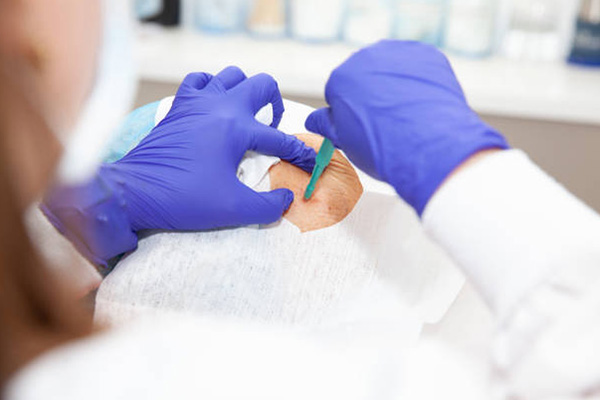
    • Fine-Needle Aspiration Cytology (FNAC): A thin needle collects cellular material from palpable or image-guided deep-seated lesions, such as thyroid nodules, breast lumps, lymph nodes, and salivary glands.

Clinical Importance and Key Benefits

Cytopathological testing is an indispensable tool in modern healthcare due to several distinct advantages:

  • Minimally Invasive: Sampling procedures require minimal recovery time and cause little to no patient discomfort compared to surgical biopsies.
  • Rapid Turnaround: Processing cell samples takes significantly less time than tissue processing, providing clinicians with fast diagnostic results.
  • Early Cancer Detection: It enables the identification of cellular atypia and dysplastic changes before tumors grow into advanced, symptomatic stages.
  • Cost-Effective Screening: Mass screening programs, such as the Pap smear, have dramatically reduced cervical cancer mortality rates worldwide.

The Diagnostic Process

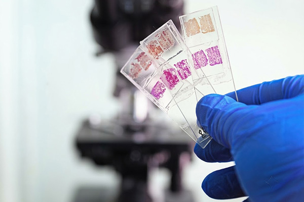
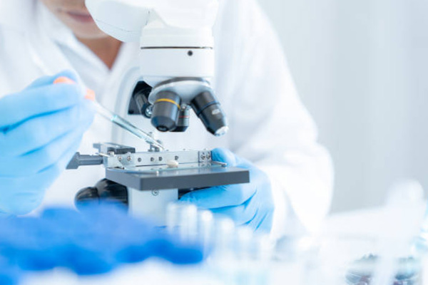
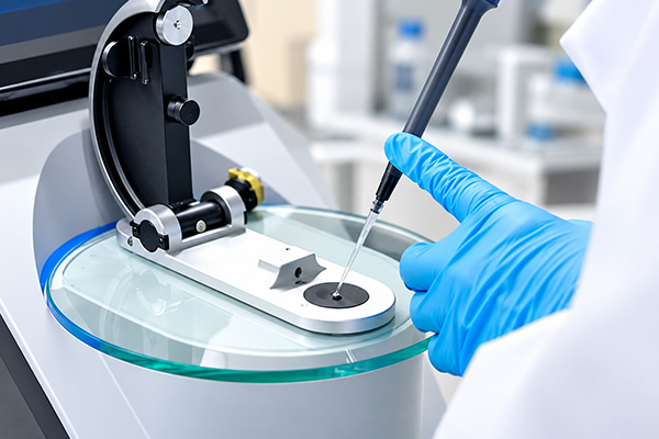
Once cells are collected, they are spread onto glass slides or prepared using Liquid-Based Cytology (LBC) techniques. LBC helps remove background debris, blood, and mucus, yielding a cleaner monolayer of cells. The slides are then stained—most commonly using the Papanicolaou (Pap) stain or Giemsa stain—to highlight cellular architecture, nuclear detail, and cytoplasmic features. Under high-magnification microscopy, the pathologist evaluates:

  • Nuclear-to-Cytoplasmic (N:C) Ratio: An enlarged nucleus often indicates malignancy.
  • Chromatin Distribution: Clumped or irregular nuclear patterns signal abnormal cell activity.
  • Cellular Arrangement: Presence of cohesive clusters, single isolated cells, or background necrosis.

When standard staining yields ambiguous results, cytopathologists utilize advanced ancillary techniques such as Immunocytochemistry (ICC) or Molecular Diagnostics (e.g., PCR and FISH) on cell blocks to pinpoint precise tumor origins and genetic mutations.

Conclusion

Cytopathology serves as the frontline defense in modern diagnostic medicine. By bridging the gap between non-invasive screening and definitive tissue biopsies, it empowers healthcare providers to diagnose conditions earlier, monitor treatment responses effectively, and improve overall patient outcomes.